Rosacea Subtypes: Demodex Mite Mapping, Azelaic Acid & Vascular Triggers
Rosacea is not simply "sensitive skin that flushes easily." It is a chronic neuro-inflammatory and vascular disorder rooted in innate immune dysregulation, microscopic mite proliferation, and abnormal cathelicidin peptide cleavage.
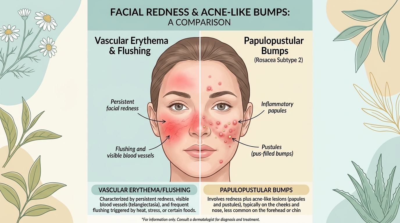
1. The 4 Rosacea Subtypes: Clinical Phenotypes
Modern clinical dermatology classifies rosacea across four distinct phenotypes based on morphological features:
Characterized by prolonged (greater than 10 minutes) central facial flushing in response to emotional, thermal, or chemical triggers. Over time, persistent microvascular dilation leads to visible, fine spider veins (telangiectasias) across the nose and malar eminences. Skin feels tight, hot, and stings readily with cosmetics.
Presents as dome-shaped erythematous papules and tiny, sterile pustules clustered centrally on the cheeks, forehead, and chin. Crucially, it lacks comedones (no blackheads or whiteheads), differentiating it from acne vulgaris. Strongly correlated with microscopic Demodex mite overgrowth.
Marked by progressive sebaceous gland hyperplasia and fibrotic dermal thickening, most commonly affecting the nasal cartilages (rhinophyma). Primarily affects men over age 40.
Involves chronic blepharitis, meibomian gland dysfunction, conjunctival hyperemia, and foreign-body sensation ("grittiness" in the eyes). Over 50% of cutaneous rosacea patients exhibit concurrent ocular symptoms.
For related muzzle rash differentials, explore our guide on Perioral Dermatitis Zero-Therapy.
2. The Demodex Parasitology & The Cathelicidin LL-37 Cascade
Recent histological investigations have illuminated the molecular engine driving Subtype 2 papules: the symbiotic relationship between Demodex folliculorum mites and human innate immunity.
In rosacea-affected facial skin, Demodex mite density reaches 10 to 15 mites per square centimeter (compared to < 1 per cm² in normal skin). When mites die, their chitinous exoskeletons and internal bacteria (Bacillus oleronius) leak into the follicular infundibulum:
- 1. Bacterial antigens activate Toll-Like Receptor 2 (TLR-2) on adjacent keratinocytes.
- 2. Activated TLR-2 triggers the abnormal overexpression of a serine protease enzyme: Kallikrein-5 (KLK-5).
- 3. Kallikrein-5 cleaves native cutaneous cathelicidin protein into its abnormal, inflammatory peptide fragment: Cathelicidin LL-37.
- 4. Cathelicidin LL-37 stimulates endothelial cells to induce rapid capillary angiogenesis (telangiectasias) while recruiting neutrophils to form inflammatory pustules.
3. Azelaic Acid Pharmacology: Why 15% Gel Outperforms 20% Cream
Azelaic acid (1,7-heptanedicarboxylic acid) is the premier evidence-based topical therapy for papulopustular rosacea. Its unique biochemical power is threefold:
- Serine Protease Inhibition: Directly inhibits Kallikrein-5 transcription, halting the cleavage of cathelicidin into inflammatory LL-37.
- Reactive Oxygen Species Scavenging: Neutralizes free radicals generated by infiltrating neutrophils.
- Anti-Demodex Activity: Suppresses follicular bacterial metabolism without inducing antibiotic resistance.
When rebuilding the compromised barrier after active rosacea flares, integrate botanical triterpenes as outlined in Centella Asiatica & Asiaticoside Science.
Frequently Asked Questions: Rosacea Science
What role do Demodex folliculorum mites play in papulopustular rosacea?
Demodex folliculorum and Demodex brevis mites reside naturally in human pilosebaceous units. In patients with papulopustular rosacea, Demodex density is up to 15 times higher than normal. When the mites die, they release endosymbiotic bacteria (such as Bacillus oleronius) that activate Toll-like receptor 2 (TLR-2) on keratinocytes, triggering kallikrein-5 and producing inflammatory cathelicidin LL-37 peptides that manifest as red papules and pustules.
Why is 15% Azelaic Acid gel considered more bioavailable than 20% cream?
Pharmacokinetic studies demonstrate that azelaic acid formulated in an aqueous gel vehicle at 15% concentration achieves up to 8 times higher cutaneous bioavailability into epidermal tissue than a 20% emulsion cream. The lipophilic emulsion base of 20% creams traps the dicarboxylic acid molecules, whereas the aqueous gel matrix drives rapid thermodynamic partitioning directly into the follicular infundibulum.
How do you differentiate between neurovascular rosacea and adult acne?
The primary clinical differentiator is the presence of open and closed comedones (blackheads and whiteheads). Acne vulgaris is intrinsically comedonal, caused by hyperkeratinization and sebum plugs. Rosacea is non-comedonal: it presents as vascular erythema, telangiectasias, and sterile inflammatory papulopustules without true sebum plugs, accompanied by facial flushing triggered by heat, spicy foods, or emotional stress.