Topical Ectoin: Cellular Protection from Dehydration Stress & Extremolyte Biology
In severe barrier dysfunction, ordinary humectants like hyaluronic acid and glycerin often falter. When ambient humidity drops or stratum corneum lipid lamellae are stripped, conventional hygroscopic agents can inadvertently draw water from viable basal layers into dry desert air. Enter ectoin: a revolutionary extremolyte that stabilizes cellular architecture through physical water structuring rather than simple passive moisture binding.
1. What Is an Extremolyte? Bacterial Osmoadaptation in Saline Deserts
Life thrives in the most inhospitable environments on Earth—from the scorching salt flats of the Wadi El Natrun desert in Egypt to hypersaline alkaline lakes. Organisms dwelling in these extreme biomes are known as extremophiles. To survive osmotic pressure that would instantaneously lyse ordinary mammalian cells, halophilic bacteria such as Halomonas elongata synthesize protective cyclic amino acid derivatives classified as extremolytes or compatible solutes.
The predominant extremolyte discovered in 1985 by Galinski and colleagues is ectoin (chemically: (S)-2-methyl-1,4,5,6-tetrahydropyrimidine-4-carboxylic acid). Within halophilic bacteria, ectoin is biosynthesized via a conserved three-step metabolic pathway encoded by the ectABC gene cluster:
- Transamination (EctB – L-DABA Aminotransferase): Converts L-aspartate-β-semialdehyde into L-2,4-diaminobutyric acid (DABA) using L-glutamate as the amino-group donor.
- Acetylation (EctA – DABA Acetyltransferase): Transfers an acetyl moiety from Acetyl-CoA to form N-γ-acetyl-2,4-diaminobutyric acid (ADABA).
- Cyclic Condensation (EctC – Ectoine Synthase): Catalyzes intramolecular condensation and dehydration of ADABA into cyclic, zwitterionic ectoin.
Because ectoin carries a negatively charged carboxylate group and a delocalized positive charge across its cyclic pyrimidine ring, it remains net neutrally charged (zwitterionic) at physiological cutaneous pH (4.8–5.5). Crucially, compatible solutes do not interfere with enzymatic processes or cellular metabolic machinery—making them remarkably non-toxic and cytoprotective when applied topically to human skin.
Learn how extremolyte cellular defense anchors broader recovery in The Reactive Skin Barrier Rehabilitation Protocol.
2. Biophysical Mechanism: Preferential Exclusion & The "Ectoin Hydro Complex"
How does a bacterial osmolyte protect human keratinocytes and stratum corneum lipid bilayers from harsh environmental degradation? In molecular biophysics, this is explained by the preferential exclusion model (Timasheff's thermodynamic framework).
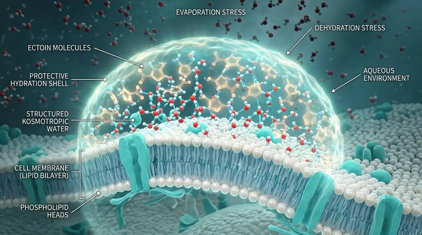
Unlike chemical moisturizers or penetration enhancers that bind directly to membrane receptors or protein backbones, ectoin is strongly kosmotropic (water-ordering). Ectoin exhibits an unfavorable thermodynamic interaction with macromolecular surfaces; it prefers to interact with bulk water molecules. Consequently, ectoin molecules are preferentially excluded from the immediate surface of proteins, corneodesmosomes, and cell membranes:
The Ectoin Hydro Complex
By being excluded from macromolecular boundaries, ectoin pulls bulk water into a rigid, highly organized 3D tetrahedral network. This generates a dense "hydration shell" surrounding adjacent cell membranes and proteins, shielding them from osmotic shrinkage and surfactant cleavage.
Lipid Bilayer Fluidity Regulation
In stratum corneum lipid lamellae, ectoin elevates the phase-transition temperature. It prevents dehydration-induced crystallization of polar headgroups, preserving the supple liquid-ordered state of ceramides, cholesterol, and free fatty acids under severe dry stress.
Because protein denaturation requires an expansion of molecular surface area, the preferential exclusion of ectoin imposes an enormous thermodynamic penalty on unfolding. Enzymes, structural keratins, and membrane transport proteins remain locked in their native, folded, functional conformations even under extreme thermal, chemical, or evaporative stress.
3. Cutaneous Cytoprotection: Langerhans Cells, HSP70 & Pollution Defense
In dermatology, the clinical value of ectoin extends far beyond standard moisture retention. It functions as an active cellular shield across three critical epidermal axes:
A. Preservation of Epidermal Langerhans Cells
Langerhans cells are the specialized dendritic antigen-presenting immune sentinels resident in the stratum spinosum. Following ultraviolet exposure (UVA and UVB) or chemical surfactant washouts, Langerhans cells undergo severe oxidative apoptosis and deplete rapidly, leaving skin immunologically vulnerable. In vivo biopsy trials demonstrate that pre-treatment with 2% ectoin prevents up to 100% of UV-induced Langerhans cell depletion, sustaining vital cutaneous immune surveillance.
B. Heat Shock Protein (HSP70) Modulation
Under environmental distress, skin cells overexpress Heat Shock Protein 70 (HSP70) as an emergency chaperone to refold damaged proteins. Because ectoin’s hydration shell physically preserves protein stability, cellular stress signals are dramatically attenuated. HSP70 expression is initiated more rapidly but at significantly lower, controlled amplitudes, sparing keratinocytes metabolic exhaustion.
C. Anti-Pollution (PM2.5) & ICAM-1 Downregulation
Airborne particulate matter (PM2.5 and PM10) binds polycyclic aromatic hydrocarbons that stimulate intracellular reactive oxygen species (ROS) and upregulate Intercellular Adhesion Molecule-1 (ICAM-1). Topical ectoin blocks particle-induced mitochondrial oxidative stress, downregulates ICAM-1 expression on endothelial and epidermal cells, and inhibits inflammatory leukocyte extravasation.
For patients whose barrier compromise is paired with microvascular hypersensitivity, explore our active-layering guidance in Rosacea Subtypes & Azelaic Acid Protocols.
4. Clinical Evidence: TEWL Kinetics & Formulating Concentrations
Unlike synthetic silicones that create temporary artificial occlusive films without altering epidermal physiology, ectoin stimulates lasting, intrinsic barrier recovery:
- Surfactant Challenge Protection: In randomized human trials using repeated sodium dodecyl sulfate (SDS) washouts, pre-treatment with topical ectoin reduced surfactant-induced transepidermal water loss (TEWL) elevation by up to 40%, preserving stratum corneum integrity against harsh cleansing agents.
- Dose-Dependent Corneometry: In cohorts with severe xerosis and atopic dermatitis, a 5.0% ectoin emulsion produced a 15% sustained reduction in TEWL, a 30% decrease in cutaneous erythema, and a 30% elevation in stratum corneum hydration within 7 days.
- Synergistic Compatibility: Ectoin does not decompose under oxidative or thermal stress and possesses a neutral zwitterionic charge. It blends seamlessly alongside gentle chemical exfoliants like polyhydroxy acids. Review the molecular advantages in our comparative analysis: Gluconolactone PHA vs Salicylic Acid.
Clinical Concentration Guidelines for Formulators
Frequently Asked Questions: Topical Ectoin Science
What is an extremolyte, and how is ectoin biosynthesized?
Extremolytes are low-molecular-weight compatible organic solutes synthesized by extremophilic microorganisms to survive extreme osmotic desiccation, intense salinity, and temperature fluctuations. Ectoin (1,4,5,6-tetrahydro-2-methyl-4-pyrimidinecarboxylic acid) is biosynthesized by halophilic bacteria such as Halomonas elongata via a conserved three-step enzymatic cascade encoded by the ectABC gene cluster: transamination of L-aspartate semialdehyde to DABA (EctB), acetylation to ADABA (EctA), and cyclic condensation catalyzed by ectoine synthase (EctC).
How does the 'preferential exclusion' model explain ectoin's hydration mechanism?
Unlike conventional humectants that bind directly to protein surfaces, ectoin acts as a strongly kosmotropic (water-structuring) zwitterion that is excluded from the immediate protein-water interface. This negative preferential interaction parameter forces surrounding water molecules into a highly structured, dense hydrogen-bonded hydration shell (the Ectoin Hydro Complex) around cell membranes and native proteins, raising the thermodynamic penalty for protein denaturation and membrane disruption.
How does ectoin protect epidermal Langerhans cells from UV damage?
Epidermal Langerhans cells—the primary antigen-presenting immune sentinels of the skin—are critically depleted following ultraviolet (UVA/UVB) irradiation. Topical ectoin prevents this depletion by stabilizing cellular membrane fluidity, limiting lipid peroxidation, and suppressing premature stress signaling, thereby preserving cutaneous immune surveillance and preventing UV-induced systemic immunosuppression.
What concentration of ectoin is optimal for compromised, reactive skin barriers?
Clinical corneometry and transepidermal water loss (TEWL) evaluations show that daily barrier repair serums achieve statistically significant hydration and cytoprotection at 0.5% to 2.0% ectoin. For severely compromised barriers, contact dermatitis, and atopic xerosis, clinical dermatitis formulations utilizing 4.0% to 7.0% ectoin demonstrate accelerated stratum corneum recovery and up to 40% attenuation of surfactant-induced barrier breakdown.