Celebrity Skin Realism: Candid Breakdown of Perioral Dermatitis, Rosacea & Barrier Fatigue
Behind 8K cinema lenses, high-output studio lighting, and multi-step cosmetic marketing lies unfiltered human biology. When prominent public figures remove thick silicone primers and candidly reveal stinging erythematous papules, the aesthetic conversation undergoes a profound clinical correction.
Clinical Overview: Deconstructing Red-Carpet Dermal Fragility
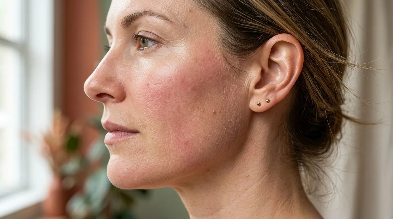
- 1. The Reality of Unfiltered Texture: High-definition cinematography and social media filters have created an artificial standard of "poreless" skin. In reality, stratum corneum micro-relief, follicular ostia, and vascular flush are physiological universals.
- 2. Perioral Dermatitis (POD) Pathogenesis: Triggered by topical corticosteroid exposure, heavy occlusive foundations, fluorinated toothpaste, and cosmetic active stacking, POD manifests as uniform 1–2mm micro-papulopustules sparing the vermilion border.
- 3. The Paradoxical Cure of Zero-Therapy: Ceasing all active serums, heavy creams, and surfactants allows endogenous lipid synthesis and acid mantle pH homeostasis to self-regulate without chemical disruption.
1. The Illusion of Poreless Perfection vs. High-Definition Reality
For over a decade, digital image post-processing, blurring algorithms, and studio-grade diffusion filters cultivated an unattainable cosmetic myth: that healthy skin is an inert, glass-like sheet devoid of topography. Yet, biological human skin is an active, living organ. It breathes through millions of pilosebaceous units, constantly undergoes microscopic desquamation, and reacts dynamically to humidity, thermal spikes, and hormonal fluctuations.
In recent years, an overdue cultural shift toward Skin Realism has gained significant momentum. High-profile figures have pulled back the curtain on production-induced skin distress. Model and entrepreneur Hailey Bieber publicly took to TikTok and interview platforms without makeup or flattering filters to document her recurring struggles with perioral dermatitis, detailing how unpredictable red rashes and sensitive bumps emerge around her nasolabial folds and chin. Similarly, actress Florence Pugh has repeatedly celebrated her real skin texture, refusing to hide hormonal breakouts or facial erythema on major red carpets, while Kendall Jenner has candidly recounted the intense emotional vulnerability of walking award shows during severe acne flares.
Modern high-definition cinema sensors (such as RED Monstro 8K and ARRI Alexa LF) capture resolutions exceeding 35 megapixels with extreme dynamic range. Under these optics, heavy pancake makeup and occlusive dimethicone-based smoothing primers do not make pores disappear; instead, they cake within the superficial skin folds (sulci cutis), highlighting dehydration cracks and micro-comedones. True dermal health on camera is not about masking texture, but restoring barrier integrity, epidermal hydration, and cellular lipid coherence.
When consumers attempt to replicate this airbrushed illusion at home by layering 10 to 12 active products—stacking glycolic acid cleansers with high-strength retinoids, direct Vitamin C, and dense occlusive balms—the outcome is rarely porcelain skin. More frequently, it triggers a catastrophic cascade of stratum corneum barrier fatigue, chronic neurovascular flushing, and inflammatory dermatoses.
2. Deconstructing Perioral Dermatitis (POD): Etiology & Triggers
Perioral dermatitis (POD)—often categorized within the broader spectrum of periorificial dermatitis when it extends to the periocular or perinasal regions—is an erythematous papulopustular disorder predominantly affecting women aged 18 to 45. Clinically documented in authoritative dermatological literature (StatPearls / NCBI Books, NBK525968), POD presents as an eruption of uniform, discrete, 1 to 2 millimeter inflammatory papules and micro-pustules on an erythematous base.
A definitive clinical hallmark of perioral dermatitis is the clear zone of sparing immediately encircling the vermilion border of the lips. While the perioral skin, chin, and nasolabial creases erupt with tight, burning, stinging papules, a distinct 2-to-3-millimeter rim of skin surrounding the lips remains completely unaffected.
| Diagnostic Parameter | Perioral Dermatitis (POD) | Papulopustular Rosacea | Acne Vulgaris |
|---|---|---|---|
| Primary Lesion Morphology | Uniform 1–2mm micro-papules & pustules; scale | Dome-shaped inflammatory papules; no comedones | Open & closed comedones (blackheads/whiteheads), cysts |
| Characteristic Distribution | Perioral, nasolabial folds, chin; vermilion border spared | Central face: convexities of cheeks, nose, forehead | Sebaceous areas: forehead, jawline, back, chest |
| Subjective Sensations | Intense burning, stinging, tightness; rarely itching | Hot vasomotor flushing, burning, thermal hypersensitivity | Occasional tenderness or pain on deep palpation |
| Comedones Present? | Strictly Absent | Strictly Absent | Always Present (Pathognomonic) |
| Response to Corticosteroids | Transient calm → Catastrophic Rebound | Exacerbates steroid rosacea | Can trigger steroid acne (uniform pustules) |
The Corticosteroid Trap & Rebound Pathogenesis
The single most devastating clinical mistake made by reactive-skin patients is reaching for an over-the-counter 1% hydrocortisone cream. Because corticosteroids act as potent vasoconstrictors and suppress local cytokine activity, the rash initially vanishes within 24 to 48 hours. However, prolonged or repeated topical steroid application paralyzes cutaneous immune surveillance, degrades intercellular collagen, and alters follicular microflora.
Upon ceasing the steroid, a violent inflammatory rebound ensues: erythema surges, capillary dilation explodes, and hundreds of pustules erupt across the lower face. Breaking this cycle requires absolute corticosteroid cessation and structured barrier stabilization.
Cosmetic Overuse, Slugging, and Fluorinated Exogenous Compounds
Beyond steroids, botanical and cosmetic chemists recognize a modern phenomenon: cosmetic barrier fatigue. The human stratum corneum possesses a tightly regulated lipid matrix consisting of ceramides, cholesterol, and free fatty acids organized into lamellar bilayers. When individuals engage in viral beauty trends such as "slugging" (applying pure petrolatum or dense waxes over existing active flares), heavy occlusives trap sweat, epidermal secretions, and follicular lipids against an already inflamed epidermis. This creates a warm, macerated anaerobic micro-environment that encourages micro-organism proliferation.
Furthermore, dental products containing high concentrations of sodium fluoride or sodium lauryl sulfate (SLS) are recognized contact irritants that continuously bathe the chin and mouth during twice-daily brushing, provoking or perpetuating perioral flares. Switching to an SLS-free, unflavored paste is a critical non-negotiable step in reactive barrier rehabilitation.
3. Rosacea, Studio Heat & The Neurovascular Axis
While perioral dermatitis clusters tightly around facial orifices, erythematotelangiectatic and papulopustular rosacea are characterized by diffuse, symmetric central facial redness, persistent telangiectasias, and profound neurovascular reactivity. In actors, red-carpet attendees, and stage performers, rosacea presents a severe occupational challenge due to the inescapable thermal environment of professional production.
Peer-reviewed biophysical research (PMC10821660; PMC4561631) demonstrates that rosacea-prone skin exhibits dysregulated transient receptor potential (TRP) channels, specifically TRPV1 and TRPA1. These thermoreceptors, located directly on sensory C-nerve fibers surrounding dermal micro-vessels, possess a lowered activation threshold.
Thermal Vasomotor Spikes
Under powerful studio key lights (which emit significant infrared radiation) or inside pressurized venue environments, cutaneous temperature rapidly rises above 36°C. TRPV1 channels open, prompting the immediate release of vasoactive neuropeptides including Substance P and Calcitonin Gene-Related Peptide (CGRP). This triggers massive arteriolar dilation, resulting in the characteristic burning, thumping facial flush.
Demodex Folliculorum Ecology
In papulopustular rosacea, microscopic saprophytic mites (Demodex folliculorum and Demodex brevis) inhabit the pilosebaceous units in densities significantly higher than in healthy controls (JEADV, doi:10.1111/jdv.70112). When heavy occlusive foundations and sweat trap sebum, mite proliferation triggers toll-like receptor 2 (TLR2) cascades, synthesizing inflammatory cytokines and generating pustules.
Elevated Transepidermal Water Loss (TEWL) is another proven pathophysiological reality in rosacea. Compromised stratum corneum lipids allow vital moisture to evaporate continuously into the dry air of film stages and soundstages. As a result, the skin behaves paradoxically: it feels excessively dry and tight, yet breaks out in inflammatory pustules when emollient creams are applied.
4. The Clinical Protocol of Zero-Therapy ("Nulltherapie")
In modern consumer skincare, the reflex response to an emerging skin flare is to apply another product: an acne spot treatment, a calming serum, a botanical oil, or a thick recovery balm. In perioral dermatitis, this interventionist mindset is precisely what perpetuates the disease.
Dermatologists and botanical chemists champion an uncompromising, clinically proven intervention: Zero-Therapy (known historically in European dermatology as Nulltherapie). Zero-therapy is the total cessation of all topical cosmetics, moisturizers, active serums, toners, sunscreens, and foaming cleansers.
The 21-Day Zero-Therapy Physiological Recovery Timeline
The skin feels extraordinarily tight, parched, and sandpaper-like. Papules may crust over and dry out. Because all synthetic humectants and emollients have been removed, the stratum corneum experiences acute superficial dryness. This is the physiological shock phase; do not surrender to the urge to apply moisturizer. Wash exclusively with lukewarm distilled or thermal spring water; gently pat dry with a clean, unbleached microfiber towel.
Erythema begins to recede from a fiery crimson to a dull salmon pink. Flaking increases as damaged, hyperkeratotic corneocytes shed naturally without chemical exfoliation. Without surfactant delipidation or emulsifiers disturbing the surface, epidermal lamellar bodies resume endogenous synthesis of sphingolipids and free fatty acids.
The surface pH re-stabilizes in its optimal acidic range (pH 4.8 to 5.5). The burning sensation upon contact with plain water disappears, signaling that sensory C-nerve fibers have re-submerged beneath a functional lipid barrier shield. Papules have flattened, leaving only transient macular erythema.
When Zero-Therapy Requires Medical Prescription Escalation
In severe cases—especially those with profound corticosteroid withdrawal rebound—zero-therapy must be paired with dermatological pharmaceutical intervention. In a landmark randomized, double-blind, vehicle-controlled clinical trial (Schwarz et al., JAAD, PMID: 18462835), topical 1% pimecrolimus cream demonstrated rapid and significant efficacy in resolving perioral dermatitis lesions without inducing cutaneous thinning, telangiectasia, or steroid rebound.
As non-steroidal topical calcineurin inhibitors (TCIs), pimecrolimus and tacrolimus selectively block T-cell activation and inhibit the synthesis of pro-inflammatory cytokines (IL-2, IFN-gamma). Other clinical interventions include prescription 15% azelaic acid gel (which provides antimicrobial, anti-keratinizing, and anti-inflammatory benefits) and low-dose oral tetracyclines (such as sub-antimicrobial doxycycline 40mg once daily), which down-regulate matrix metalloproteinases (MMPs) rather than acting primarily as an antibiotic.
5. Step-by-Step Barrier Rehabilitation for High-Stress Reactive Skin
Once the acute inflammatory flare has subsided and zero-therapy has neutralized active papules, the skin enters a vulnerable transitional state. Returning immediately to pre-flare routines or harsh foaming cleansers will reignite the dermatitis within 72 hours. Rebuilding must follow rigorous lipid biochemistry principles.
Reintroduce The Physiological 3:1:1 Equimolar Lipid Ratio
Rather than slathering pure plant oils (which often have high oleic acid profiles that destabilize lipid packing) or single-ingredient ceramides, select clinical barrier formulations engineered with the 3:1:1 ratio: 3 parts Ceramides (NP, AP, EOP), 1 part Cholesterol, and 1 part Free Fatty Acids. Pioneered by dermatological barrier biologist Dr. Peter Elias, this exact stoichiometric proportion accelerates stratum corneum lamellar bilayer repair by over 40% compared to non-physiological emollients.
Deploy Targeted Botanical Anti-Inflammatories
Select non-comedogenic, fungal-acne-safe soothing actives with proven calming kinetics:
- • Colloidal Oat Beta-Glucan: Possesses 20% higher water-binding capacity than hyaluronic acid while suppressing NF-kB inflammatory cascades.
- • Pure Madecassoside (from Centella Asiatica): Accelerates collagen synthesis and re-epithelialization while reducing IL-1alpha production.
- • Zinc PCA & Micronized Sulfur: Provide mild antimicrobial suppression of opportunistic microflora without degrading lipid lamellae.
Institute The "Banned Ingredient Matrix" During Vulnerability
For a minimum of 60 days following flare resolution, strictly eliminate the following cosmetic components:
6. Frequently Asked Questions: Skin Realism & Perioral Dermatitis
Why does room-temperature tap water burn like acid during a dermatitis flare? ↓
When the stratum corneum lipid bilayers are stripped, naked epidermal nerve endings (unmyelinated C-fibers) are exposed directly to the outside environment. Plain tap water has a neutral or slightly alkaline pH (7.0–8.0) and zero osmotic pressure compared to cellular fluids. The resulting osmotic shock causes immediate ionic depolarization of TRPV1 receptors, which transmit severe burning signals directly to the brain.
Can I wear mineral foundation if I have an important public appearance? ↓
If an event is unavoidable, strictly avoid liquid silicone-heavy or long-wear oil-based foundations. Opt instead for pure, loose mineral powder consisting solely of pharmaceutical-grade zinc oxide, titanium dioxide, and iron oxides without bismuth oxychloride, talc, or dimethicone. Zinc oxide provides mild anti-inflammatory and physical UV protection without forming an occlusive barrier that traps heat. Remove gently using micellar water formulated with non-ionic surfactants, followed by a distilled water rinse.
How does azelaic acid work for both perioral dermatitis and rosacea? ↓
Azelaic acid is a naturally occurring dicarboxylic acid that operates through multiple distinct pathways: it inhibits the generation of reactive oxygen species (ROS) by neutrophils, down-regulates kallikrein 5 and cathelicidin LL-37 expression in rosacea, suppresses micro-bacterial proliferation in follicular units, and normalizes abnormal keratinocyte maturation without causing peeling or skin thinning. Prescription 15% gel formulations are extensively validated in clinical trials.
Is perioral dermatitis contagious to family members or partners? ↓
No. Perioral dermatitis is an inflammatory and barrier disorder, not an infectious disease. It cannot be transmitted between individuals through physical contact, shared towels, or personal items. While normal commensal skin flora (such as yeasts or bacteria) may proliferate within disrupted follicular channels, they are opportunistic secondary factors rather than communicable pathogens.
Clinical Evidence & Peer-Reviewed References
- StatPearls / NCBI Books: Tolaymat L, Hall CA. Perioral Dermatitis. StatPearls Publishing; Updated 2024. NBK525968. NCBI Bookshelf.
- Journal of the American Academy of Dermatology: Schwarz CW, et al. A randomized, double-blind, vehicle-controlled adult study of 1% pimecrolimus cream in the treatment of perioral dermatitis. J Am Acad Dermatol. 2008. PMID: 18462835.
- PMC / Clinical Rosacea Biophysics: Transient Receptor Potential (TRP) channels and neurovascular reactivity in cutaneous flushing. PMC10821660 & PMC4561631. PMC Article.
- Journal of the European Academy of Dermatology and Venereology (JEADV): Demodex folliculorum density and epidermal barrier disruption in rosacea subtypes. doi:10.1111/jdv.70112. JEADV Study.
- Frontiers in Medicine / Epidermal Biology: Stratum corneum lipid lamellae degradation from multi-step cosmetic overload and surfactants. PMC9436712. PMC Article.